
Photo-induced force microscopy
Photo-induced Force Microscopy (PiFM), Molecular Vista
The Photo-induced Force Microscope (PiFM) and Infrared Spectroscopy (PiF-IR) is a revolutionary technique that uses mechanical instead of optical detection to measure the near-field optical interaction between the cantilever tip and sample to pass beyond the Abbé diffraction limit of light.
PiFM leverages local polarization of the sample by tip-enhanced optical illumination, resulting in highly localized forces acting between the AFM cantilever tip and the sample surface which are easily detected by atomic force microscopy (AFM). Coupled to a mid-infrared (MIR) light source, it can perform nanoscale Infrared Spectroscopy (nano-IR) that allows for simultaneous acquisition of 3D topographic data with molecular chemical information at high spatial resolution (~ 5 nm) and excellent spectral resolution (~ 1 cm−1).
In PiFM, the tip-sample interface generates two types of short-range forces: (1) the usual van der Waals (vdW) force that non-contact AFM (NC-AFM) relies on for mapping the topography of the sample surface, and (2) the photo-induced force (PiF) generated by near field illumination. Both forces act over a very short range, coming into play only when the tip is atomically close to the sample surface (~10 nm or less).
PiFM utilizes two vibrational modes of the rectangular AFM cantilever to measure the two forces independently – the interatomic vdW force with one mode, and the photo-induced force (PiF) with another. Since the vibration of the two modes is completely independent, motion in one mode generally does not influence the other.
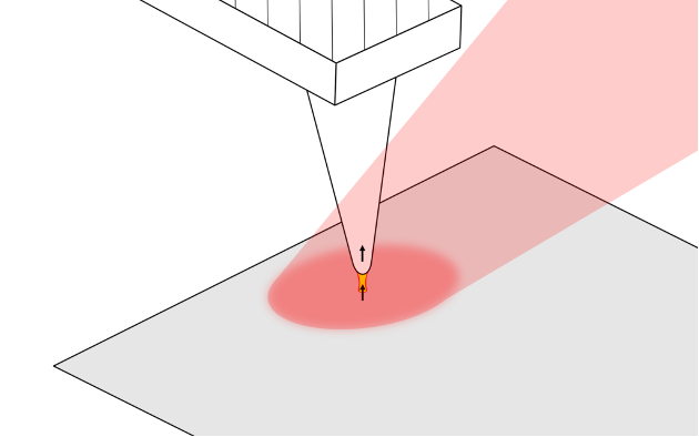
The metal-coated AFM tip creates a highly local enhanced field (yellow) from the PiF excitation laser (red). This field locally polarizes the sample, resulting in tip-sample forces that are detected by PiFM.
Instrumentation
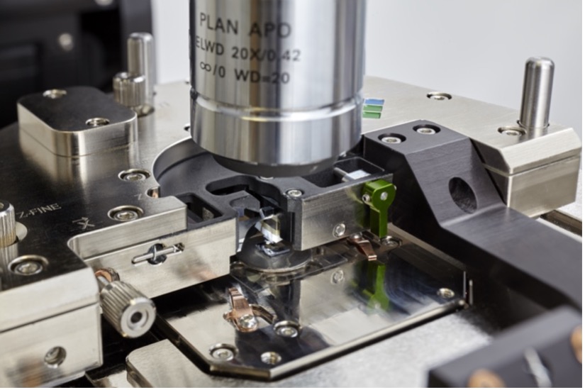
PiFM Vista One at RSES is an original PiF microscope manufactured by Molecular Vista (San José, USA) for nano‑IR chemical analysis. The instrument acquires nanoscale chemical maps and point spectra at higher detail than FTIR or nano-FTIR. It features mono-layer molecular sensitivity (2-30nm depth) and can characterize organic and inorganic materials at 5 nm spatial resolution and 1 cm-1 spectral resolution.
Sub-10 nm PiF‑IR spectroscopy
- 1 wavenumber spectral resolution with a widely tuneable laser for nano-IR experiments
- Tunable IR lasers can sweep a full PiF‑IR spectrum in as little as 100 ms.
- Use existing FTIR spectra to identify materials.
Sub-10 nm PiFM chemical mapping
- Fixed-wavelength PiFM images can be acquired in minutes with a range of up to 100 µm in X and Y.
- hyPIR™ imaging and our automated principle component analysis tools provide complete image and spectral data‑sets with minimal effort.

Sample formats
- Sample Size: less than 50 mm x 50 mm
- Sample Thickness: less than 10 mm
- Sample Shape: PiFM is based on AFM. If AFM measurement cannot be done on the sample, then PiFM measurement cannot be done either. Therefore, it may be very much worthwhile to try the sample with an AFM to ensure that the region of interest is accessible by AFM.
- Reference Samples: When providing reference samples for analysis of blended samples or for identification of potential contaminants, please provide enough material for both ATR-FTIR measurements and PiFM measurements. These measurements are possible for films if the thickness is over ~ 500 nm. Please use a substrate that is not too conductive (glass, silicon, mica, sapphire, etc. are good choices for substrates).
- Sample Storage/Transport: PiFM is surface sensitive enough to detect monolayer of molecules. When shipping, do not use gelpak sample containers as they will outgas and contaminate the sample surface. Use Fluoroware if available. If not, use wafer carrier trays made out of Natural Polypropylene as they have lower outgassing [1]. If adhesive needs to be used, please use metal tapes with acrylic adhesive.
Data Analysis Software
- SurfaceWorks 3.0
Access
The instrument is accessible to all at cost-recovery basis. It is available as a fee-for-service or for supervised in-person usage after training by the lab manager.
- Contact lab manager to discuss options
- Prerequisites for in-person users: Completion of ANU Pulse Online Training Module Laser Safety and WHS induction plus additional user training through the lab manager where required.
Acknowledge this facility
Please add the following statement to the Acknowledgment of your manuscripts and Theses:
"We acknowledge the ANU NanoPhase Facility for access to its instrumentation and lab manager Dr Kathryn Hayward for her support with PiFM analyses."
PiFM Publications:
Förster, M. W., Prelević, D., Chen, C., & Shcheka, S. S. (2026). Lower crustal differentiation and transient shallow assembly of the Laacher See magma system. Lithos 544-545, 108769.
Vo, P. H., Vogel, C., Clark, S. M., Bishop, D. P., Droas, M., Partyka-Jankowska, E., Förster, M. W., Adam. C., Strezov, V., & Nguyen, H. T. (2026). PFAS released from the metals recovery process of the lithium-ion battery black mass. Journal of Hazardous Materials 509, 141978.
Förster, M.W., Otter, L.M., Brocks, J.J., Jayasoma, K., Cisneros-Lazaro, D., Nowak, D., Stolarski, J. and Knowles, B., 2025. Quality Control Measures for Enhancing Confidence in Nanoscale IR spectroscopy. Geostandards and Geoanalytical Research 49, 685-703.
Foley, S.F., Ezad, I.S., Shu, C. and Förster, M.W., 2025. Melting of amphibole-apatite-rich metasomes in the continental mantle and comparison of melt compositions with natural igneous rocks. Lithos, 500-501, 107976.
Förster, M.W., Otter, L.M., Brocks, J.J., Jayasoma, K., Cisneros-Lazaro, D., Nowak, D., Stolarski, J. and Knowles, B., 2024. A PiFM spectral library for the identification of molecular contaminants in geochemical laboratories, Mendeley Data, V1, doi: 10.17632/zmxssj8w38.1
Förster, M.W., Chen, C., Foley, S.F., Alard, O. and Yaxley, G.M., 2024. Fluid loss to the fore-arc controls the recycling efficiency of nitrogen in subduction zones. Chemical Geology, 650, 121985.
Ezad, I.S., Saunders, M., Shcheka, S.S., Fiorentini, M.L., Gorojovsky, L.R., Förster, M.W. and Foley, S.F., 2024. Incipient carbonate melting drives metal and sulfur mobilization in the mantle. Science Advances, 10(12), p.eadk5979
Otter, L.M., Eder, K., Kilburn, M.R., Yang, L., O’Reilly, P., Nowak, D.B., Cairney, J.M. and Jacob, D.E., 2023. Growth dynamics and amorphous-to-crystalline phase transformation in natural nacre. Nature Communications, 14(1), 2254.
Ezad, I.S., Shcheka, S.S., Buhre, S., Buhre, A., Gorojovsky, L.R., Shea, J.J., Förster, M.W. and Foley, S.F., 2023. Rapid quench piston cylinder apparatus: An improved design for the recovery of volatile-rich geological glasses from experiments at 0.5–2.5 GPa. Review of Scientific Instruments, 94(5) 055107
Otter, L.M., Förster, M.W., Belousova, E., O’Reilly, P., Nowak, D., Park, S., Clark, S., Foley, S.F. and Jacob, D.E., 2021. Nanoscale chemical imaging by photo‐induced force microscopy: technical aspects and application to the geosciences. Geostandards and Geoanalytical Research, 45(1), 5-27.
