X-ray fluorescence (XRF) scanning
The ANU Research School of Earth Sciences operates a cutting-edge X-ray Fluorescence (XRF) scanning facility, capable of non-destructive elemental analysis from magnesium to uranium on a variety of samples including sediment cores and rocks.
Groups
Content navigation
About
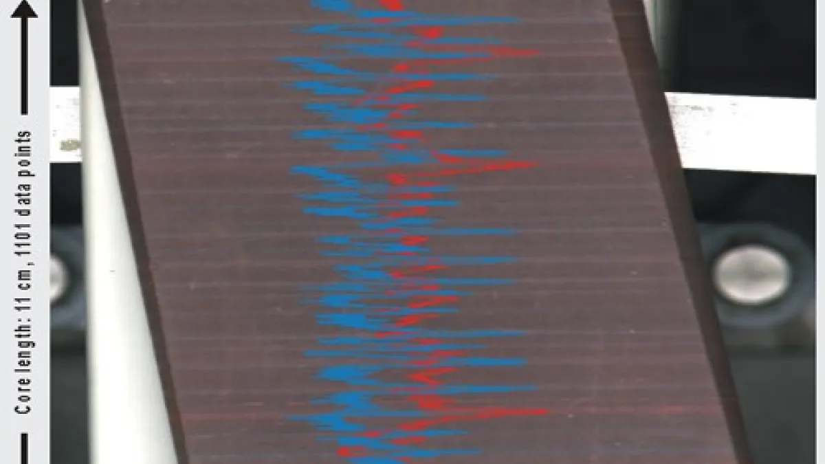
The Ocean and Climate Change group, led by Prof. Eelco Rohling, operate a state-of-the-art XRF scanning facility at RSES. The Avaatech core-scanner performs non-destructive analysis of elements from Mg through to U, on split sediment cores and smoothed surfaces of stalagmites, corals, rocks, etc, as well as on discrete samples. Down-core resolution is 10 mm to 0.1 mm, with a 15 to 1 mm beam-width.
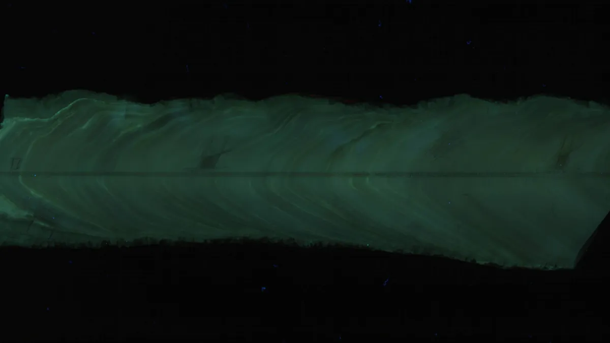
The scanner is also equipped with a digital Colour Line camera that records images with visible or UV light. This allows high‐resolution images to be exactly cross‐referenced with the elemental scans. It also means that a path can be defined in any direction across the sample surface simply by clicking the mouse on the core photo, and then a series of XRF measurements are taken along that path.
Location
ANU


